Neural tissue interfaces
Discovering the impact of insertion parameters for optimizing neural yield,
biocompatibility and quality of multi-channel extracellular recordings

Strategies for seamless
probe implantation and integrationOptimizing parameters: insertion speed, probe angle, site layout, shank geometry, novel probe materials, coatings, vasoconstrictors, artefact filtering and more.
High-density neural probes allow the recording of brain electrical activity with high spatial and temporal resolution. These electrical imaging devices if implanted chronically, can be used for the long-term monitoring of the activity of single neurons to study brain mechanisms where electrophysiological changes over time are relevant (e.g. learning and memory). Furthermore, neural electrical imaging and stimulation techniques are important components of several neural prosthetic device and brain-computer interface applications.

Unfortunately, the long-term performance of most neural probes is limited, the electrical impulses (or spikes) of neurons can be recorded for days or weeks, sometimes for months, but rarely for longer periods. The scale of performance degradation may depend on numerous factors like the biocompatibility of the implanted device, the biological response of the brain tissue around the implantation site, hemorrhage by rupturing blood vessels during insertion and micro- or macromovements of the neural probe relative to the brain. These factors can significantly contribute to the damage and subsequent death of neurons and to an elevated tissue reaction resulting in the formation of a glial scar around the implanted device.

Our aim is the development of a complex implantation strategy which may contribute to achieve high-quality neural recordings for long periods of time. This strategy includes several methods to minimize, on the one hand, the tissue damage during probe insertion and on the other hand, the biological response of the brain tissue to the implanted probes. Such methods, for instance, are the local cooling of the brain tissue near the implant site, local application of vasoconstrictor substances to decrease probe insertion-related bleeding, design and 3D printing of protective components (e.g. head caps) for chronically implanted probes, or a careful design of the insertion conditions (e.g. using a very slow insertion speed).
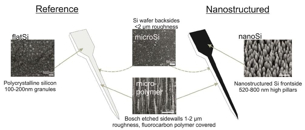
Furthermore, we are investigating novel probe materials and coatings which may enhance the biocompatibility of the implanted devices which in turn may provide better quality neural signals and for a longer term.